
Understanding Spinal Stenosis
Spinal stenosis occurs when spaces within the spine narrow, potentially compressing the spinal cord and nerves. This constriction can lead to pain, numbness, and functional limitations.
The spinal cord acts as a vital communication pathway, transmitting messages between the brain and the body. Disorders can disrupt these signals, impacting movement and organ function.
Vertebrae, the individual bones of the spine, stack to form a protective column. These 33 bones, including fused sections, safeguard the delicate spinal cord within.
The spinal canal is the passageway through which the spinal cord travels. Its size can vary, and narrowing can contribute to stenosis, causing nerve compression and related symptoms.
What is Spinal Stenosis?
Spinal stenosis isn’t a disease itself, but rather a condition characterized by the narrowing of spaces within your spine. This narrowing can put pressure on the spinal cord and the nerves that branch off from it. While it can occur anywhere along the spine – cervical, thoracic, or lumbar – lumbar spinal stenosis is the most common.
This narrowing isn’t always present from birth; it often develops over time due to degenerative changes associated with aging. These changes can include osteoarthritis, thickening of ligaments, and the formation of bone spurs. The result is a reduced space for the spinal cord and nerves, leading to a variety of symptoms.
Symptoms can range from mild pain and numbness to more severe issues like muscle weakness and difficulty with walking. The specific symptoms depend on the location and severity of the stenosis. Understanding the underlying cause and the specific areas affected is crucial for developing an effective management plan, often incorporating targeted exercises.
The Spinal Cord and its Function
The spinal cord is a long, cylindrical bundle of nerves extending from the base of the brain down the back. It’s the primary pathway for communication between the brain and the rest of the body, essentially acting as a superhighway for vital signals.
This crucial structure transmits sensory information – like touch, pain, and temperature – to the brain, and motor commands from the brain to muscles and organs. Without a properly functioning spinal cord, voluntary movement, reflexes, and even organ function would be severely compromised.
Nerves branch out from the spinal cord through openings in the vertebrae, forming a complex network that reaches every part of the body. When the spinal cord or these nerves are compressed, as in spinal stenosis, it disrupts these signals, leading to a range of neurological symptoms. Maintaining spinal cord health is therefore paramount for overall well-being.
Vertebrae: The Building Blocks of the Spine
The human spine is comprised of 33 individual bones called vertebrae, though two of these sections – the sacrum and coccyx – are fused together. These bones aren’t simply stacked; they are intricately designed and interconnected to provide both stability and flexibility.
Each vertebra features a central body, a vertebral arch, and several processes (like the lamina and transverse processes) that serve as attachment points for muscles and ligaments. This complex structure allows for a wide range of motion while protecting the delicate spinal cord that runs through the vertebral canal.
The vertebrae are separated by intervertebral discs, which act as shock absorbers and further contribute to spinal flexibility. Understanding the structure of the vertebrae is crucial when discussing conditions like spinal stenosis, where changes to these bones can contribute to nerve compression and pain.
Spinal Canal: Protecting the Spinal Cord
The spinal canal is the hollow passageway formed by the vertebral bodies, providing crucial protection for the spinal cord and its nerve roots. This bony tunnel extends from the base of the skull down to the lower back, acting as a robust shield against injury.
Within the spinal canal, the spinal cord is suspended in cerebrospinal fluid, which further cushions and nourishes this vital structure. Openings, called foramina, along the sides of the spinal canal allow nerve roots to exit and branch out to the rest of the body, enabling movement and sensation.
The size of the spinal canal can vary between individuals; some are born with a naturally narrower canal, predisposing them to spinal stenosis. Narrowing of the spinal canal, whether congenital or developed over time, can compress the spinal cord and nerves, leading to a range of symptoms.

Causes of Spinal Stenosis
Spinal stenosis arises from various factors, including congenital narrowing, degenerative changes, facet joint issues, and herniated discs—all impacting the spinal canal’s space.
Congenital Spinal Stenosis
Congenital spinal stenosis refers to a narrowing of the spinal canal present at birth. Some individuals are simply born with a smaller spinal canal than others, predisposing them to potential nerve compression later in life. This isn’t typically caused by an injury or disease process, but rather a developmental variation.
While symptoms may not appear immediately, they often develop during adulthood as age-related changes in the spine occur. These changes, such as thickening of ligaments or the formation of bone spurs, can further reduce the space within the spinal canal, exacerbating the congenital narrowing.
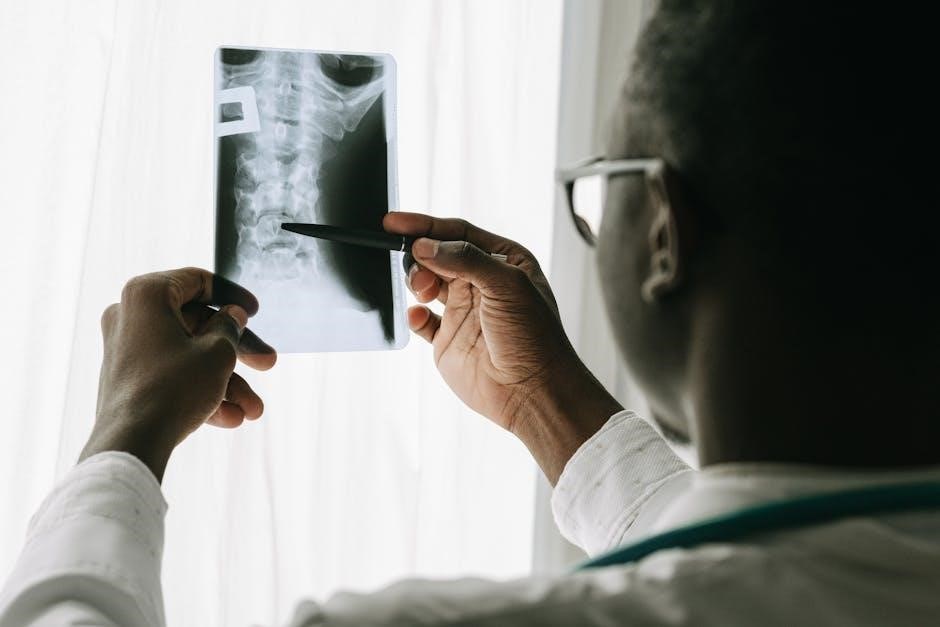
Diagnosis usually involves imaging techniques like MRI or CT scans to visualize the spinal canal’s dimensions. Management focuses on alleviating symptoms and preventing progression, often involving physical therapy and, in some cases, surgical intervention to decompress the spinal cord. Early identification and proactive management are key for individuals with congenital spinal stenosis.

Degenerative Changes & Spinal Stenosis
Degenerative spinal stenosis is the most common form, developing over time due to age-related wear and tear on the spine. As we age, the ligaments within the spine can thicken, and bone spurs may form, gradually narrowing the spinal canal. These changes reduce the space available for the spinal cord and nerves.
Osteoarthritis, a common joint condition, plays a significant role. The facet joints, which provide stability to the spine, can become arthritic, leading to inflammation and bone growth. Degenerative disc disease, where the intervertebral discs lose hydration and height, also contributes to narrowing.
These degenerative processes often occur gradually, with symptoms developing slowly over years. Common symptoms include pain, numbness, and weakness in the legs and feet. Management typically involves conservative treatments like physical therapy, pain medication, and lifestyle modifications. In severe cases, surgical decompression may be considered to relieve pressure on the spinal cord.
Facet Joint Issues and Stenosis
Facet joints are crucial for spinal stability and movement, located at the posterior aspect of each vertebra. When these joints become inflamed or arthritic, they can contribute significantly to spinal stenosis. Hypertrophy, or overgrowth, of the facet joints is a common finding in individuals with stenosis, narrowing the spinal canal and potentially compressing nerve roots.
This overgrowth often results from osteoarthritis, a degenerative joint disease. As cartilage breaks down, the body attempts to repair the damage, leading to bone spur formation. These bone spurs encroach upon the spinal canal, exacerbating stenosis. Inflammation within the facet joints can also cause pain and contribute to nerve irritation.
Addressing facet joint issues is often a key component of managing spinal stenosis. Treatment options range from conservative measures like physical therapy and pain medication to more invasive procedures like facet joint injections or, in severe cases, surgical decompression to create more space for the nerves.
Herniated Discs & Their Role
Intervertebral discs act as cushions between the vertebrae, providing shock absorption and allowing for spinal flexibility. A herniated disc occurs when the soft, gel-like center of a disc protrudes through a tear in the tougher outer layer. This protrusion can significantly contribute to spinal stenosis by directly compressing the spinal cord or nerve roots.
While not always the primary cause of stenosis, a herniated disc can exacerbate existing narrowing of the spinal canal. The displaced disc material can physically impinge on neural structures, leading to pain, numbness, and weakness in the areas served by those nerves. The location of the herniation – cervical, thoracic, or lumbar – dictates the specific symptoms experienced.
Managing a herniated disc alongside spinal stenosis often involves a combination of approaches, including pain management, physical therapy focused on core stabilization, and, in some cases, surgical intervention to relieve pressure on the spinal cord and nerves. Careful assessment is crucial to determine the best course of action.
Exercises for Spinal Stenosis: General Principles
Exercise programs for spinal stenosis prioritize core strength, low-impact movements, and avoiding positions that worsen symptoms. Gradual progression is key for safety and effectiveness.
Importance of Core Strengthening
Core muscles play a crucial role in supporting the spine and maintaining proper posture. Strengthening these muscles – including abdominals, back muscles, and pelvic floor – can significantly alleviate symptoms associated with spinal stenosis. A strong core acts as a natural brace, reducing stress on the spine and improving stability during movement.
Exercises targeting the core don’t necessarily need to be complex. Simple movements like pelvic tilts, abdominal bracing, and bird-dog exercises can be highly effective. These exercises help improve spinal alignment and reduce the load on compressed nerves. Consistent core work can also enhance balance and coordination, minimizing the risk of falls and further injury.
However, it’s vital to perform core exercises with proper form to avoid exacerbating symptoms. Individuals with spinal stenosis should consult with a healthcare professional or physical therapist to develop a personalized core strengthening program tailored to their specific needs and limitations.
Low-Impact Exercise Focus
Low-impact exercises are paramount when managing spinal stenosis, as they minimize stress on the spine while promoting mobility and strength. High-impact activities, like running or jumping, can aggravate symptoms by increasing compression on the spinal cord and nerves. Instead, focus on exercises that are gentle on the joints and spine.
Excellent low-impact options include walking, swimming, water aerobics, and cycling on a stationary bike. These activities provide cardiovascular benefits without placing excessive strain on the back. Water-based exercises are particularly beneficial, as the buoyancy reduces body weight and pressure on the spine.
Remember to start slowly and gradually increase the intensity and duration of your workouts. Pay close attention to your body and stop if you experience any pain or discomfort. A tailored exercise plan, developed with guidance from a healthcare professional, is crucial for maximizing benefits and minimizing risks.
Avoiding Aggravating Movements
Identifying and avoiding movements that worsen spinal stenosis symptoms is crucial for effective management. Certain positions and activities can increase pressure on the spinal cord and nerves, leading to pain, numbness, or weakness. Common aggravating movements often involve excessive bending backward (extension), twisting, or prolonged sitting.
Individuals with spinal stenosis may find relief by modifying daily activities. For example, using proper lifting techniques – bending at the knees and keeping the back straight – can minimize strain. Avoiding prolonged static postures, like sitting for extended periods, is also beneficial. Regular changes in position and short walks can help alleviate pressure.
Listen carefully to your body and note which movements consistently trigger symptoms. A physical therapist can provide personalized guidance on posture correction and activity modification to help you navigate daily life with greater comfort and functionality. Prioritizing these adjustments is key to preventing symptom flare-ups.
Specific Exercises for Spinal Stenosis (PDF Focus)
PDF resources often detail McKenzie, extension, flexion (with caution), and lateral flexion exercises. These targeted movements aim to decompress the spine and alleviate stenosis symptoms.
McKenzie Exercises for Spinal Stenosis
McKenzie exercises, frequently detailed in downloadable PDF guides, are a cornerstone of non-surgical management for spinal stenosis. These exercises primarily focus on repeated spinal extension movements, aiming to centralize pain – meaning moving pain from the extremities towards the spine, indicating a reduction in nerve compression.
A common starting point is the prone press-up. Lying face down, the individual gradually pushes up onto their forearms, holding the extended position for a short duration. This is repeated several times. Another key exercise is the standing back extension, involving gentle backward bending of the upper back.
PDF resources emphasize the importance of performing these exercises slowly and deliberately, paying close attention to any changes in symptoms. It’s crucial to stop if pain worsens or radiates further down the legs. These exercises aren’t a one-size-fits-all solution, and proper guidance from a healthcare professional, often outlined within the PDF instructions, is essential for safe and effective implementation.
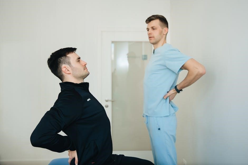
Extension Exercises
Extension exercises, often detailed in comprehensive PDF guides for spinal stenosis, aim to create more space within the spinal canal by gently arching the back. These movements can help relieve pressure on the spinal cord and nerves, reducing pain and improving function. Many PDFs highlight the importance of controlled movements and proper form.
Examples include prone lying (lying on your stomach) and back extensions, performed either on the floor or using a specialized extension bench. Standing backbends, performed cautiously, are also common. These exercises encourage a posterior shift of the vertebrae, potentially widening the spinal canal.
PDF resources consistently advise starting slowly and gradually increasing the range of motion as tolerated. It’s vital to monitor symptoms closely; any increase in leg pain or neurological symptoms warrants immediate cessation and consultation with a healthcare provider. The downloadable PDFs often include illustrations and detailed instructions to ensure correct execution and maximize benefits.
Flexion Exercises (Caution Advised)
Flexion exercises – movements that round the back – are often approached with caution in spinal stenosis management, as detailed in many PDF exercise guides. While some individuals may experience relief, flexion can exacerbate symptoms by further narrowing the spinal canal in certain cases. Therefore, careful consideration and professional guidance are crucial.
PDF resources typically emphasize a trial-and-error approach, starting with very gentle movements like chin tucks or seated forward bends (performed minimally). It’s essential to monitor for any increase in leg pain, numbness, or weakness. If symptoms worsen, flexion exercises should be discontinued immediately.
The downloadable PDFs often include specific warnings and modifications for individuals with different types and locations of spinal stenosis. Core engagement during flexion is also stressed to provide spinal support. Remember, these exercises aren’t universally beneficial and require individualized assessment and supervision by a qualified healthcare professional.
Lateral Flexion Exercises
Lateral flexion exercises, involving bending sideways, are frequently included in spinal stenosis exercise PDFs, but with specific instructions for safe execution. These movements aim to improve spinal mobility and potentially relieve nerve compression, though effects vary greatly between individuals.
PDF guides often demonstrate seated or standing side bends, emphasizing slow, controlled movements. It’s crucial to avoid forcing the bend and to stop if any pain radiates down the legs. Maintaining a neutral spine and engaging core muscles during the exercise is also highlighted.
Many resources caution against overdoing lateral flexion, particularly if it increases neurological symptoms. Gentle stretches, holding the bend for a few seconds, are generally recommended over repetitive, forceful movements. The downloadable materials often suggest combining lateral flexion with other exercises for a more comprehensive approach to managing spinal stenosis.
Exercises Targeting Specific Spinal Regions
PDF exercise guides detail region-specific movements for stenosis – cervical, thoracic, and lumbar. These tailored routines address unique anatomical needs and symptom presentations for optimal relief.
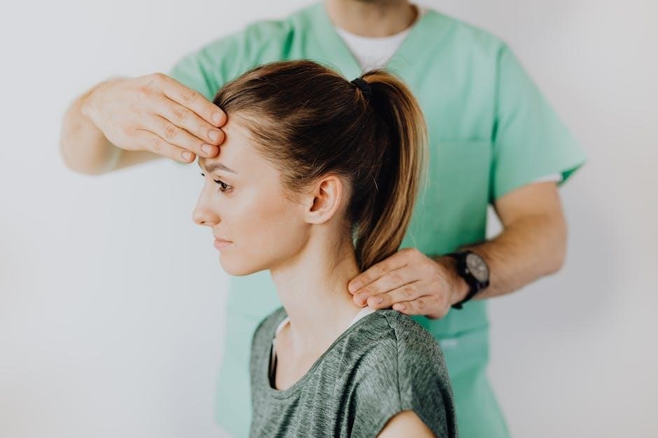
Cervical Spinal Stenosis Exercises
PDF resources often showcase gentle exercises for cervical spinal stenosis, focusing on restoring neck mobility and reducing nerve compression. Chin tucks are frequently recommended, strengthening deep neck flexors and improving posture. These exercises gently retract the head, creating more space within the cervical canal.
Neck extensions, performed cautiously, can also provide relief by opening the spaces between vertebrae. However, avoid forceful movements or hyper-extension, which could exacerbate symptoms. Shoulder blade squeezes improve posture and support the neck. Lateral neck stretches gently increase range of motion, easing muscle tension.
Many PDF guides emphasize the importance of slow, controlled movements and stopping immediately if pain increases. It’s crucial to consult a healthcare professional before starting any new exercise program, ensuring the exercises are appropriate for your specific condition and severity of stenosis. Proper form is paramount to avoid further injury.
Thoracic Spinal Stenosis Exercises
PDF exercise guides for thoracic spinal stenosis typically prioritize improving spinal mobility and core stability. Cat-cow stretches are commonly recommended, gently articulating the spine and promoting flexibility. These movements help to decompress the thoracic region and relieve pressure on nerves.
Thoracic rotations, performed seated or lying down, enhance rotational range of motion. Wall angels strengthen the muscles supporting the upper back and improve posture. Gentle back extensions, using a foam roller, can also provide relief, but should be approached cautiously.
Core strengthening exercises, like pelvic tilts and bird-dogs, are vital for supporting the thoracic spine. PDFs often stress the importance of avoiding excessive bending or twisting, which can aggravate symptoms. Always consult a healthcare professional before beginning any exercise regimen, ensuring it’s tailored to your individual needs and limitations.
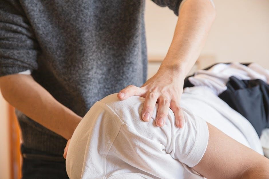
Lumbar Spinal Stenosis Exercises
PDF resources for lumbar spinal stenosis exercises frequently emphasize decompression techniques. McKenzie exercises, particularly prone press-ups, are often featured, aiming to centralize pain by extending the lumbar spine. These exercises can help reduce nerve compression and improve walking distance.
Pelvic tilts and abdominal bracing strengthen core muscles, providing support to the lower back. Hamstring stretches, performed gently, improve flexibility and reduce tension on the spine. Water exercises, like walking in a pool, offer low-impact relief.
PDF guides consistently advise against exercises that involve forward bending or twisting, as these can exacerbate symptoms. Bird-dog exercises improve core stability without stressing the lumbar spine. Remember to consult a healthcare professional before starting any new exercise program, ensuring proper form and avoiding aggravation.

Important Considerations & Safety
PDF guides stress consulting a healthcare professional before starting exercises. Listen to your body, modifying or stopping if pain increases. Gradual progression is key for safety.
Consulting a Healthcare Professional
Before initiating any exercise program for spinal stenosis, a consultation with a healthcare professional is paramount. This isn’t merely a suggestion, but a crucial step towards ensuring safety and maximizing benefits. A doctor, physical therapist, or qualified healthcare provider can accurately diagnose the specific type and location of your stenosis.
They will assess your overall health, identify any contraindications, and tailor an exercise plan to your individual needs and limitations. PDF resources often emphasize this point, but they cannot replace personalized medical advice. A professional can demonstrate proper form, preventing potential injury and ensuring exercises are performed effectively.
Furthermore, they can help differentiate between acceptable discomfort and pain that signals a problem. They can also monitor your progress and adjust the program as needed. Self-treating based solely on PDF guides carries inherent risks; professional guidance is essential for a safe and successful outcome.
Listening to Your Body
A cornerstone of any successful exercise regimen for spinal stenosis is attentive self-monitoring. While PDF exercise guides can provide valuable routines, they cannot account for your unique response. Pay close attention to how your body feels during and after each exercise. Sharp, radiating pain is a clear signal to stop immediately.
Dull aches or mild discomfort may be acceptable, but should not increase in intensity; Don’t push through pain, believing it’s simply “working out the kinks.” PDF resources often caution against aggravating movements, but your body is the ultimate guide. Recognize the difference between muscle soreness and nerve-related pain.
Modify exercises as needed, reducing range of motion or repetitions. Rest when you need to, and don’t hesitate to consult your healthcare professional if symptoms worsen or persist. Prioritize gentle, controlled movements and respect your body’s limitations. Ignoring warning signs can exacerbate your condition.
Progression and Modification of Exercises
PDF exercise guides for spinal stenosis often present a starting point, not a rigid endpoint. Gradual progression is key to building strength and flexibility without exacerbating symptoms. Begin with a reduced range of motion and fewer repetitions than suggested, focusing on proper form.
As your strength improves and pain subsides, incrementally increase repetitions or hold times. Modifications are crucial; if an exercise causes discomfort, alter it. For example, reduce the depth of a stretch or perform the exercise seated instead of standing. PDFs may offer variations, but don’t be afraid to create your own.
Listen to your body and avoid pushing through pain. Progression should be slow and steady, guided by your individual response. Regularly reassess your tolerance and adjust the program accordingly. Consulting with a physical therapist can ensure safe and effective progression tailored to your specific needs and condition.
Resources for Spinal Stenosis Information (PDFs)
Numerous organizations offer downloadable PDF exercise guides for spinal stenosis. The National Institute of Neurological Disorders and Stroke (NINDS) provides comprehensive information on spinal cord disorders, often linking to relevant resources. Hospital websites, like those of major medical centers, frequently host patient education materials, including exercise protocols.
The American Academy of Orthopaedic Surgeons (AAOS) offers patient-focused PDFs detailing spinal stenosis and potential treatments, sometimes including basic exercise illustrations. Searching for “spinal stenosis exercises PDF” yields results from physical therapy clinics and rehabilitation centers, offering varied programs.
However, exercise caution when using online resources. Always prioritize PDFs from reputable sources and consult with a healthcare professional before starting any new exercise regimen. These resources are supplementary and should not replace personalized medical advice. Verify the date of publication to ensure the information is current.