The EKG Detective‚ a monthly column‚ emphasizes deductive logic for EKG analysis‚ starting today‚ 04/09/2026 at 01:12:02․
What is an EKG and Why is it Important?
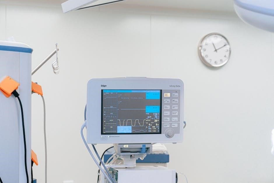
An Electrocardiogram (EKG or ECG) is a non-invasive diagnostic tool crucial for evaluating the heart’s electrical activity․ It graphically records these electrical signals‚ providing valuable insights into heart rate‚ rhythm‚ and potential abnormalities․ Understanding EKGs is fundamental for healthcare professionals‚ and readily available basic EKG PDF guides are excellent starting points for learning․
The importance of an EKG lies in its ability to detect a wide range of cardiac conditions‚ from subtle arrhythmias to life-threatening myocardial infarctions (heart attacks); Early detection‚ facilitated by EKG interpretation skills‚ allows for prompt intervention and improved patient outcomes․ As highlighted by “The EKG Detective” column launching on 04/09/2026‚ a logical approach to EKG analysis is key․
Furthermore‚ mastering EKG interpretation‚ aided by resources like introductory basic EKG PDF materials‚ empowers clinicians to make informed decisions regarding patient care‚ treatment plans‚ and monitoring strategies․ It’s a cornerstone of cardiology and emergency medicine․
Understanding the Electrical System of the Heart

The heart’s rhythmic beating is governed by a sophisticated electrical conduction system․ This system initiates impulses in the Sinoatrial (SA) node‚ the heart’s natural pacemaker‚ then travels through the atria‚ to the Atrioventricular (AV) node‚ and finally down the Bundle of His and Purkinje fibers‚ causing ventricular contraction․ An EKG captures this electrical activity․
Understanding this pathway is vital for interpreting EKG waveforms․ Disruptions at any point – from the SA node to the Purkinje fibers – can manifest as abnormalities on the EKG tracing․ Resources like a basic EKG PDF can visually illustrate this system‚ aiding comprehension․
As “The EKG Detective” column‚ beginning 04/09/2026‚ will demonstrate‚ a systematic approach‚ coupled with knowledge of the heart’s electrical physiology‚ is essential․ Studying a basic EKG PDF alongside learning the conduction system provides a strong foundation for accurate interpretation․

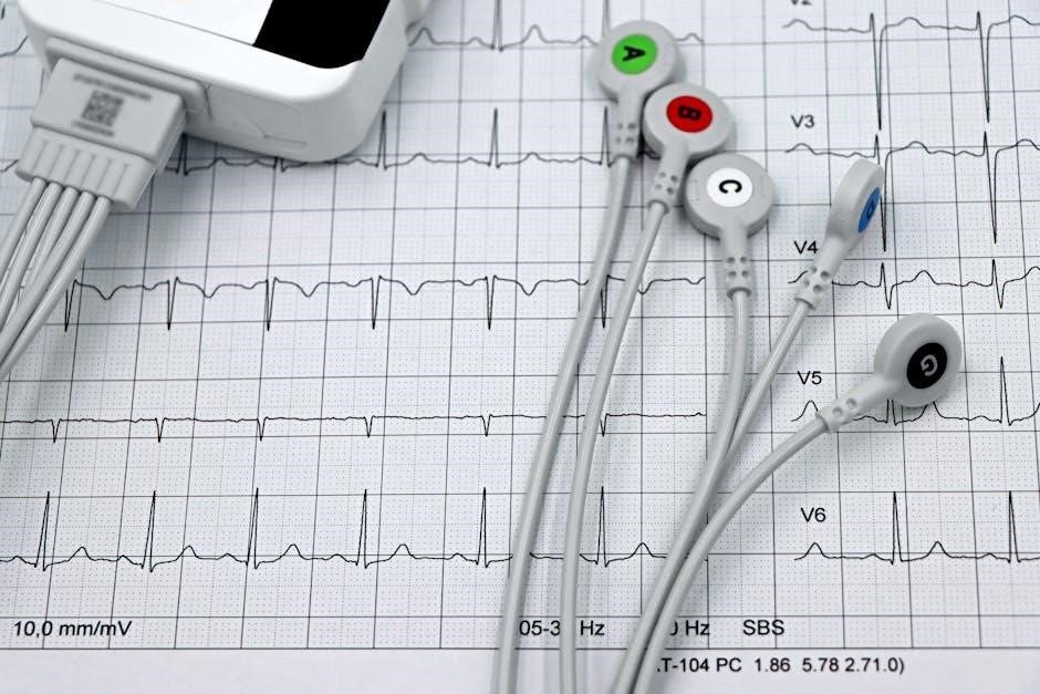
EKG Components and Waveforms
EKG tracings display P waves‚ QRS complexes‚ and T waves; a basic EKG PDF visually details each component’s significance for analysis․
P Wave: Atrial Depolarization
Understanding the P wave is fundamental in basic EKG interpretation․ Representing atrial depolarization‚ this initial upward deflection signifies the electrical activation of the atria․ A basic EKG PDF guide will visually demonstrate a normal P wave’s characteristics: typically small‚ rounded‚ and upright in most leads․
Analyzing the P wave’s morphology – its shape‚ duration‚ and amplitude – provides crucial diagnostic clues․ Abnormal P waves can indicate atrial enlargement‚ atrial ischemia‚ or ectopic atrial rhythms․ For instance‚ a peaked P wave might suggest right atrial enlargement‚ while a notched P wave could indicate left atrial enlargement․
A basic EKG PDF resource will often include examples of normal and abnormal P waves‚ aiding in accurate identification․ Consistent practice with these visual aids is key to mastering P wave interpretation and building a solid foundation in EKG analysis․ Recognizing P wave abnormalities is a critical step in identifying various cardiac conditions․
QRS Complex: Ventricular Depolarization
The QRS complex represents ventricular depolarization – the electrical activation of the ventricles‚ triggering their contraction․ A basic EKG PDF will illustrate this as the sharp‚ typically positive deflection following the P wave․ Understanding its components – the Q wave‚ R wave‚ and S wave – is vital․
A normal QRS duration is generally between 0․06 and 0․10 seconds․ Prolonged QRS complexes suggest delayed ventricular activation‚ potentially due to bundle branch blocks or ventricular pre-excitation․ Conversely‚ a narrow QRS complex indicates normal ventricular conduction․
Basic EKG PDF guides often showcase examples of varying QRS morphologies and durations․ Learning to accurately measure the QRS duration and identify abnormal QRS patterns is crucial for diagnosing arrhythmias and ventricular conduction abnormalities․ Mastering QRS complex interpretation forms a cornerstone of effective EKG analysis․
T Wave: Ventricular Repolarization
The T wave signifies ventricular repolarization – the return of the ventricles to their resting state‚ preparing for the next contraction․ A basic EKG PDF will visually demonstrate this as a typically rounded‚ asymmetrical waveform following the QRS complex․ Its polarity is usually positive in most leads‚ though exceptions exist․
T wave abnormalities‚ such as inversion or flattening‚ can indicate ischemia‚ injury‚ or electrolyte imbalances․ Peaked T waves may suggest hyperkalemia․ Analyzing T wave morphology alongside other EKG components is essential for comprehensive interpretation․
Basic EKG PDF resources frequently detail common T wave variations and their clinical significance․ Accurate T wave assessment requires understanding its relationship to the QRS complex and the overall rhythm․ Mastering T wave interpretation is vital for identifying potentially life-threatening cardiac conditions․
PR Interval: Assessing AV Conduction
The PR interval represents the time it takes for the electrical impulse to travel from the atria to the ventricles‚ reflecting atrioventricular (AV) conduction․ A basic EKG PDF will illustrate measuring it from the beginning of the P wave to the start of the QRS complex․
A normal PR interval typically ranges from 0․12 to 0․20 seconds․ Prolongation suggests a first-degree AV block‚ indicating delayed conduction․ Shortening can signify pre-excitation syndromes like Wolff-Parkinson-White․
Basic EKG PDF guides emphasize the importance of consistent PR interval measurement․ Variations can signal underlying cardiac issues․ Accurate assessment requires identifying the precise start and end points on the EKG tracing․ Understanding the PR interval is crucial for diagnosing AV conduction abnormalities and guiding appropriate clinical management․
QT Interval: Total Ventricular Activity
The QT interval represents the total time for ventricular depolarization and repolarization․ A basic EKG PDF will demonstrate how to measure it from the beginning of the QRS complex to the end of the T wave․ Correct QT interval assessment is vital‚ as abnormalities can indicate increased risk of dangerous arrhythmias․

Normal QT intervals are rate-dependent and are often corrected for heart rate using formulas like Bazett’s (QTc)․ Prolonged QTc intervals can predispose individuals to torsades de pointes‚ a life-threatening ventricular tachycardia․ Shortened QT intervals are less common but can also be associated with arrhythmias․
Basic EKG PDF resources highlight the importance of accurate measurement and correction․ Factors like electrolyte imbalances and medications can affect the QT interval․ Careful interpretation‚ alongside clinical context‚ is essential for patient safety․

Analyzing EKG Rhythms
EKG Detective columns utilize deductive logic for rhythm interpretation; basic EKG PDF guides aid in identifying normal sinus rhythms and common arrhythmias efficiently․

Normal Sinus Rhythm
Understanding the Baseline: A normal sinus rhythm (NSR) represents the heart’s typical electrical activity‚ originating from the sinoatrial (SA) node – the heart’s natural pacemaker․ Utilizing a basic EKG PDF resource is crucial for beginners to visually recognize NSR characteristics․ Key features include consistent P waves preceding each QRS complex‚ a regular R-R interval (the time between ventricular depolarizations)‚ and a heart rate generally between 60 and 100 beats per minute․
PDF Guide Application: Basic EKG PDF guides often dedicate sections to NSR‚ illustrating ideal waveforms and providing practice strips for identification․ These guides emphasize the importance of assessing rhythm regularity and the relationship between atrial and ventricular activity․ The EKG Detective approach‚ focusing on deductive reasoning‚ complements PDF study by encouraging systematic waveform analysis․ Consistent practice with these resources builds confidence in recognizing a healthy heart rhythm and forms a foundation for identifying abnormalities․
Mastering NSR identification is the first step in EKG interpretation‚ and a solid basic EKG PDF is an invaluable tool for achieving this proficiency․
Common Arrhythmias: Bradycardia & Tachycardia
Beyond Normal: Bradycardia (slow heart rate – below 60 bpm) and tachycardia (fast heart rate – above 100 bpm) represent deviations from normal sinus rhythm․ A basic EKG PDF is essential for differentiating these arrhythmias and understanding their underlying causes․ These guides typically dedicate chapters to common arrhythmias‚ providing illustrative EKG strips and detailed explanations․
PDF-Guided Analysis: When using a basic EKG PDF‚ focus on rate calculation and rhythm regularity․ Bradycardia often presents with prolonged R-R intervals‚ while tachycardia shows shortened intervals․ The EKG Detective’s deductive approach encourages identifying the rate first‚ then analyzing the waveform morphology․ Understanding P wave presence and relationship to QRS complexes is vital․
Practical Application: Basic EKG PDF resources often include case studies of bradycardia and tachycardia‚ allowing for practical application of learned concepts․ Recognizing these common arrhythmias is a fundamental skill in EKG interpretation‚ and consistent study with reliable PDF guides is key․
Atrial Fibrillation and Atrial Flutter
Irregular Heartbeats: Atrial fibrillation (AFib) and atrial flutter are common arrhythmias characterized by rapid‚ irregular atrial activity․ A basic EKG PDF is invaluable for recognizing these patterns‚ often showcasing characteristic EKG features․ These PDFs typically dedicate sections to differentiating AFib from atrial flutter‚ highlighting key waveform differences․
PDF-Focused Identification: In AFib‚ the baseline often appears wavy with absent P waves․ Atrial flutter displays “sawtooth” waves․ A basic EKG PDF will illustrate these patterns clearly․ The EKG Detective’s method stresses systematic waveform analysis‚ starting with the P wave (or lack thereof)․ Rate control is a crucial aspect of management․
Learning Resources: Many basic EKG PDF guides include algorithms for identifying AFib and flutter‚ aiding in quick and accurate diagnosis․ Understanding these arrhythmias is essential‚ and consistent review of PDF materials reinforces recognition skills․ Practice interpreting example tracings found within these resources․
Identifying Myocardial Infarction (Heart Attack) on an EKG
EKG Detective utilizes deductive logic; a basic EKG PDF aids in recognizing STEMI versus NSTEMI patterns and MI locations effectively․
STEMI vs․ NSTEMI
Distinguishing between STEMI (ST-elevation myocardial infarction) and NSTEMI (non-ST-elevation myocardial infarction) is crucial for immediate treatment decisions․ A basic EKG PDF guide will illustrate the key differences․ STEMI presents with characteristic ST-segment elevation‚ indicating complete coronary artery occlusion and requiring urgent reperfusion therapy – either thrombolytics or percutaneous coronary intervention (PCI)․
Conversely‚ NSTEMI typically shows ST-segment depression or T-wave inversion‚ suggesting partial occlusion or significant coronary artery disease․ While still serious‚ NSTEMI often allows for a more measured approach to management‚ including medication and potential angiography․ The EKG Detective column highlights how deductive reasoning‚ coupled with a solid understanding from a basic EKG PDF‚ can clarify these distinctions․ Recognizing these patterns quickly‚ aided by visual references in a basic EKG PDF‚ directly impacts patient outcomes and treatment strategies․
EKG Changes in Different MI Locations
A basic EKG PDF is invaluable for understanding how myocardial infarction (MI) manifests differently on an electrocardiogram depending on the affected heart region․ Anterior MIs often present with ST-segment elevation in leads V1-V4‚ while inferior MIs typically show changes in leads II‚ III‚ and aVF․ Lateral MIs affect leads I‚ aVL‚ V5‚ and V6․
Posterior MIs can be subtle‚ often showing reciprocal changes in anterior leads․ Recognizing these location-specific patterns‚ as detailed in a comprehensive basic EKG PDF‚ aids in pinpointing the blocked coronary artery․ The EKG Detective emphasizes that mastering these nuances requires diligent study and practice with illustrative examples found within a quality basic EKG PDF resource․ Accurate interpretation‚ guided by a basic EKG PDF‚ is vital for appropriate and timely intervention‚ improving patient prognosis significantly․
Resources for Further Learning (Basic EKG PDF Focus)
Basic EKG PDF guides and online courses‚ like those highlighted by The EKG Detective‚ are crucial for mastering EKG interpretation skills effectively․
Recommended Basic EKG PDF Guides
Embarking on EKG interpretation requires accessible learning resources‚ and several excellent Basic EKG PDF guides are readily available․ These PDFs often provide a structured approach‚ beginning with the fundamentals of cardiac electrophysiology and progressing to waveform analysis․ Look for guides that clearly illustrate normal sinus rhythm and common arrhythmias‚ offering detailed explanations alongside representative EKG tracings․
Many freely downloadable PDFs focus on practical application‚ including step-by-step instructions for systematic EKG reading․ Prioritize guides that emphasize a deductive reasoning approach‚ mirroring the philosophy of resources like The EKG Detective column․ Consider PDFs from reputable medical institutions or cardiology societies‚ ensuring accuracy and up-to-date information․ Remember to supplement PDF study with interactive online courses and hands-on practice for optimal comprehension and skill development․ A well-chosen PDF can be an invaluable tool in your EKG learning journey․
Online EKG Interpretation Courses
While Basic EKG PDF guides offer a foundational understanding‚ online courses provide dynamic learning experiences crucial for mastering EKG interpretation․ These courses often incorporate interactive modules‚ quizzes‚ and case studies‚ reinforcing concepts presented in PDF resources․ Many platforms offer tiered learning‚ starting with basic waveform recognition and progressing to complex arrhythmia identification and myocardial infarction diagnosis․
Look for courses that align with a deductive reasoning approach‚ similar to the principles highlighted in The EKG Detective column‚ emphasizing systematic analysis․ Consider courses offering Continuing Medical Education (CME) credits for professional development․ Supplementing PDF study with online courses allows for real-time feedback and personalized learning․ Explore options from accredited institutions and reputable cardiology organizations to ensure quality and accuracy․ Combining both PDF guides and online courses provides a comprehensive and effective path to EKG proficiency․